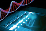
Dendra2


Green-to-red photoswitchable fluorescent protein Dendra2

- Monomer, successful performance in fusions
- Irreversible photoconversion from a green to a red fluorescent form
- High contrast of photoconversion
- Activated by UV-violet and blue light
- Matures at a wide range of temperatures
- Recommended for tracking cell, organelle, and protein movement, and for determination of protein half-life

Performance and use

Dendra2 efficiently matures both at 20°C and 37°C, which makes possible the use of the protein in wide range of experimental systems, from cultured mammalian cells to cold-blooded animals. Mammalian cells transiently transfected with Dendra2 expression vectors display an evenly distributed green signal without aggregation within 10-12 hrs after transfection. No cell toxicity is observed. High photostability of photoconverted Dendra2 (more than 3 times higher than of DsRed) makes it particularly useful for long-term protein tracking applications.

Dendra2 successful performance has been proven in many fusions including that with cytoplasmic β-actin, BH3 interacting domain death agonist (BID), nucleolar protein fibrillarin, vimentin, and α-tubulin.

Labeling of intracellular proteins with Dendra2.

Confocal images of HeLa cells transiently expressing Dendra2-tagged proteins.

High contrast of photoconversion:: In response to intense 405 nm or 488 nm light irradiation, Dendra2 undergoes irreversible photoconversion expressed in a decrease in green and appearance of red fluorescence.

Green-to-red photoconversion of Dendra2.

(A) Cell photolabeling: HEK293 Phoenix Eco cells were transiently transfected with Dendra2 gene under the control of CMV promoter. Dendra2 was converted to the red state in selected cells by brief illumination with 405-nm (left cell) or 488-nm (upper right and middle cells) lasers. Then confocal images of cells were made in green and red channels and overlaid. (B) Photoconversion in cell nucleus: HeLa cells were transiently transfected with Dendra2 gene under the control of CMV promoter. Dendra2 was converted in a nucleus by brief illumination with 405-nm laser. Then confocal images of a cell were made in green and red channels and overlaid.

After complete photoconversion, red fluorescence of Dendra2 increases more than 150-300 times, whereas the level of green fluorescence becomes more than 10-15 times lower. Thus, the increase in the red-to-green fluorescence ratio results in about a 4000-fold contrast. Considerable decrease of green fluorescence during Dendra2 photoconversion provides a molecular tool to simultaneously track both the movement of the activated protein and its replacement with the non-activated form.

Green-to-red photoconversion of Dendra2-tagged proteins.

HeLa cells were transiently transfected with vectors encoding Dendra2-tagged fusion proteins which was photoconverted in a selected region by 488-nm laser. Confocal images were made after photoconversion in green and red channels.
SEE movie

Dendra2 use for determination of protein half-life: In the method proposed, cells are transfected with a construct coding for target protein fused with a photoswitchable tag (PAFP). A steady-state concentration of the fusion protein and corresponding fluorescent signal depends on protein synthesis and maturation rates as well as protein degradation rate. After photoconversion of the photoswitchable tag in a whole cell, a pool of distinct fluorescent molecules appears, which is independent of the synthesis and maturation of the new PAFP molecules. Thus, the decay of the activated fluorescence directly corresponds to the degradation of the PAFP-tagged protein. Time-lapse imaging of the activated signal allows for quantification of degradation process in real-time at the single cell level [Zhang et al., 2007].

To test the applicability of Dendra2 for determination of protein half-life, it was fused with IkappaB-alpha protein, having well-characterized decay in cells. Cells with moderate expression levels of IkappaB-α-Dendra2 demonstrated the expected, predominantly cytoplasmic, localization of green fluorescence. After photoconversion, time-lapse series showed fast decay of the red signal with a half-life of 1.5-2 hrs. The addition of a proteasome inhibitor immediately terminated red fluorescence decay. Thus, the decrease of red fluorescent signal was caused by proteasomal degradation of the fusion protein. The rate of red signal decay was in good agreement with the available data on the half-life of IkappaB-alpha obtained using cycloheximide chase. It has been shown earlier that the phorbol ester, phorbol 12-myristate 13-acetate (PMA), increases the IkappaB-alpha degradation rate. Indeed, a considerable acceleration of red fluorescence decay after cell treatment with PMA was detected using photoactivation of IkappaB-α-Dendra2 [Zhang et al., 2007].

Green-to-red photoconversion of Dendra2-tagged proteins.

HeLa cells were transiently transfected with vectors encoding Dendra2-tagged fusion proteins which was photoconverted in a selected region by 488-nm laser. Confocal images were made after photoconversion in green and red channels.

References:

  • Zhang L, Gurskaya NG, Merzlyak EM, Staroverov DB, Mudrik NN, Samarkina ON, Vinokurov LM, Lukyanov S, Lukyanov KA. Method for real-time monitoring of protein degradation at the single cell level. Biotechniques. 2007; 42 (4):446, 448, 450. / pmid: 17489230
Copyright 2002-2008 Evrogen. All rights reserved.
Evrogen JSC, 16/10 Miklukho-Maklaya str., Moscow, Russia, +7(495)4298020, e-mail:evrogen@evrogen.com