|
Green fluorescent protein TagGFP
- Fast protein maturation
- Bright green fluorescence
- Proven availability to generate stably transfected cell lines
- Monomeric protein with successful performance in fusions
- Recommended for protein labeling
 |
TagGFP is an enhanced bright mutant of the Aequorea macrodactyla GFP-like protein [Xia et al., 2002]. TagGFP possesses bright green fluorescence with excitation/emission maxima at 482 and 505 nm, respectively. It is optimized for expression at 37oC. TagGFP is more pH-stable than EGFP.
|
Main properties
TagGFP normalized excitation (thin line) and emission (thick line) spectra.
Download TagGFP spectra (xls)
| | CHARACTERISTIC | |
|---|
* Brightness is a product of extinction coefficient and quantum yield, divided by 1000.
| | Molecular weight, kDa | 27 | | Polypeptide length, aa | 238 | | Fluorescence color | green | | Excitation maximum, nm | 482 | | Emission maximum, nm | 505 | | Quantum yield | 0.59 | | Extinction coefficient, M-1cm-1 | 58 200 | | Brightness* | 34.3 | | Brightness, % of EGFP | 104 | | pKa | 4.7 | | Structure | monomer | | Aggregation | no | | Maturation rate at 37°C | fast | | Photostability | high | | Cell toxicity | not observed | | Main advantages | bright green monomeric fluorescent protein |
|
|---|
Recommended filter sets and antibodies
The protein can be recognized using Anti-Tag(CGY)FP antibody (Cat.# AB121) or Anti-GFP antibody (Cat.# AB011) available from Evrogen.
TagGFP can be detected using common fluorescence filter sets for EGFP, FITC, and other green dyes. Recommended Omega Optical filter sets are QMAX-Green, XF100-2, XF100-3, XF115-2, and XF116-2.
Performance and use
TagGFP can be easily expressed and detected in a wide range of organisms. Mammalian cells transiently transfected with TagGFP expression vectors produce bright fluorescence in 10-12 hrs after transfection. No cell toxic effects and visible protein aggregation are observed.
Being expressed in mammalian cells, TagGFP shows brightness and maturation speed similar to those of EGFP. However, compared with EGFP, TagGFP matures more than two times faster in E. coli cells.
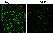 | TagGFP and EGFP expression in E. coli, 10 hrs after transformation
|
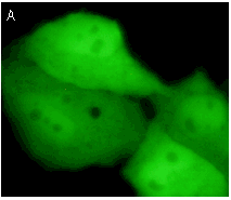
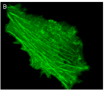
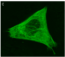
TagGFP performance in fusions has been demonstrated in the β-actin, α-tubulin and mitochondria-targeting signal models. It can be used in multicolor labeling applications with blue, true-yellow, red, and far-red fluorescent dyes.
 |  |  | |
|---|
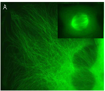
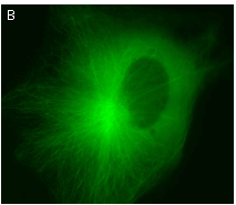
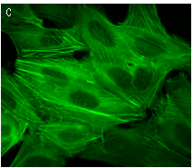
TagGFP expression in transiently transfected mammalian cells
(A) Whole-cell expression; (B) TagGFP fusion with human β-actin; (C) TagGFP fusion with human α-tubulin.
See also 3D movie of TagGFP-tagged tubulin
|
TagGFP suitability to generate stably transfected cells has been proven by Marinpharm company.
References:
-
Xia NS, Luo WX, Zhang J, Xie XY, Yang HJ, Li SW, Chen M, Ng MH.
Bioluminescence of Aequorea macrodactyla, a common jellyfish species in the East China Sea.
Mar Biotechnol (NY). 2002; 4 (2):155-62. / pmid: 14961275
|