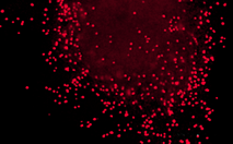
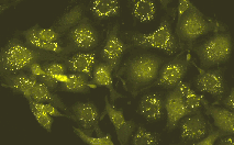
Tools for labeling of subcellular structures

- Easy labeling of cell compartments and proteins
- Wide collection of fluorescent colors
- No exogenous chemical compounds or cofactors required

Subcellular localization vectors: labeling of peroxisomes
CAT.#PRODUCTREPORTERCOLOR OF FLUORESCENCESIZEPRICE
The prices do not include delivery. The prices vary in different countries. Please contact your local distributor for exact prices and delivery information.
FP313 pmKate2-peroxi peroxisome-targeted mKate2 far-red 20 μg € 400
FP606 pPhi-Yellow-peroxi peroxisome-targeted PhiYFP-m yellow 20 μg € 400
  A
  B

Labeling of peroxisomes:

(A) – Transiently transfected HeLa cells expressing mKate2 fusion with peroxisomal targeting signal. Scale bar represents 10 μm. Image from Shcherbo et al., 2009.
(B) – Stably transfected T24 human bladder carcinoma cells expressing peroxisome-targeted PhiYFP-m. Image was kindly provided by Dr. Christian Petzelt (Marinpharm).

Copyright 2002-2011 Evrogen. All rights reserved.
Evrogen JSC, 16/10 Miklukho-Maklaya str., Moscow, Russia, Tel +7(495)988-4084, Fax +7(495)988-4085, e-mail:evrogen@evrogen.com