
|
||||||||||
|
||||||||||


TurboFP602
SUPPORTRESOURCES |
|
||||||||||||||||||||||||||||||||||||||||||||||||||||||||||||||||||||||
|
TurboFP602 is a red-shifted variant of the red fluorescent protein TurboRFP from sea anemone Entacmaea quadricolor [Merzlyak et al., 2007]. TurboFP602 possesses true-red fluorescence (with excitation/emission maxima at 574/602 nm, respectively), optimal for detection via most popular filter sets, and is easily distinguished from background signals. TurboFP602 exhibits fast maturation and high pH stability. TurboFP602 is mainly intended for applications where fast appearance of true-red fluorescence is crucial. It is specially recommended for cell and organelle labeling and for tracking the promoter activity in autofluorescent tissues. |

Main properties

TurboFP602 normalized excitation (thin line) and emission (thick line) spectra. |
| ||||||||||||||||||||||||||||||||||||||
|---|---|---|---|---|---|---|---|---|---|---|---|---|---|---|---|---|---|---|---|---|---|---|---|---|---|---|---|---|---|---|---|---|---|---|---|---|---|---|---|
Recommended filter sets and antibodies
TurboFP602 can be recognized using Anti-tRFP antibody (Cat.# AB233) available from Evrogen.
TurboFP602 can be detected using TRITC filter set or similar. Recommended Omega Optical filter sets are QMAX-Red and XF102-2.
Performance and use
TurboFP602 can be expressed and detected in a wide range of organisms. Mammalian cells transiently transfected with TurboFP602 expression vectors produce bright fluorescence in 8-12 hrs after transfection. No cytotoxic effects or visible protein aggregation are observed.
Despite its dimeric structure, TurboFP602 performs well in some fusions. However, for protein labeling applications we recommend using specially optimized monomeric TagFPs.
TurboFP602 suitability to generate stably transfected cells has been proven by Marinpharm company.
TurboFP602 can be used in multicolor labeling applications with blue, cyan, green, and yellow fluorescent dyes.
 |  |  | |
|---|---|---|---|
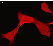
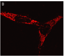
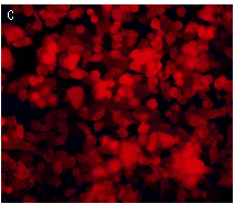
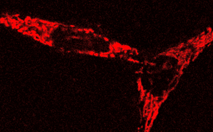
TurboFP602 expression in mammalian cells.(A) Transiently transfected Hela cells; (B) transiently transfected HeLa cells expressing mitochondria-targeted TurboFP602; (C) stably transfected human melanoma cell line Mel-Juso. Image (C) was kindly provided by Dr. Christian Petzelt (Marinpharm). | |||
| Variant | Description | Related vector | Cat.# | Click for image |
|---|---|---|---|---|
| ||||
| Humanized TurboFP602 | TurboFP602 codon usage is optimized for high expression in mammalian cells [Haas et al., 1996], but it can be successfully expressed in many other heterological systems. |
|
FP712 | |
| TurboFP602-mito fusion | A mitochondrial targeting sequence (MTS) is fused to the TurboFP602 N-terminus. MTS was derived from the subunit VIII of human cytochrome C oxidase [Rizzuto et al., 1989; Rizzuto et al., 1995]. When expressed in mammalian cells, this variant provides true-red fluorescent labeling of mitochondria. |
|
FP717 |
|
References:
- Haas J, Park EC, Seed B. Codon usage limitation in the expression of HIV-1 envelope glycoprotein. Curr Biol. 1996; 6 (3):315-24. / pmid: 8805248
- Merzlyak EM, Goedhart J, Shcherbo D, Bulina ME, Shcheglov AS, Fradkov AF, Gaintzeva A, Lukyanov KA, Lukyanov S, Gadella TW, Chudakov DM. Bright monomeric red fluorescent protein with an extended fluorescence lifetime. Nat Methods. 2007; 4 (7):555-7. / pmid: 17572680
- Rizzuto R, Brini M, Pizzo P, Murgia M, Pozzan T. Chimeric green fluorescent protein as a tool for visualizing subcellular organelles in living cells. Curr Biol. 1995; 5 (6):635-42. / pmid: 7552174
- Rizzuto R, Nakase H, Darras B, Francke U, Fabrizi GM, Mengel T, Walsh F, Kadenbach B, DiMauro S, Schon EA. A gene specifying subunit VIII of human cytochrome c oxidase is localized to chromosome 11 and is expressed in both muscle and non-muscle tissues. J Biol Chem. 1989; 264 (18):10595-600. / pmid: 2543673
|
Copyright 2002-2023 Evrogen. All rights reserved. Evrogen JSC, 16/10 Miklukho-Maklaya str., Moscow, Russia, Tel +7(495)988-4084, Fax +7(495)988-4085, e-mail:evrogen@evrogen.com |