
|
||||||||||
|
||||||||||


TurboFP650
SUPPORTRESOURCES |
|
|||||||||||||||||||||||||||||||||||||||||||||||||||||||||||||||||||||||||||
|
TurboFP650 (scientific name eqFP650) is a red-shifted variant of TurboFP635 (Katushka) [Shcherbo et al., 2010]. TurboFP650 is characterized by a strong bathochromic shift, with excitation and emission peaks at 592 nm and 650 nm, respectively. TurboFP650 demonstrates fast maturation at 37°C and a high pH-stability and photostability. The protein does not show residual short wavelength fluorescence of intermediate or alternative chromophore forms, in contrast to E2-Crimson [Strack et al., 2009], which exhibits a second bright blue emission peak, and mNeptune [Lin et al., 2009], which has a pronounced green peak. TurboFP650 is specially recommended for whole body imaging and multicolor applications. |

Main properties

TurboFP650 normalized excitation (thin line) and emission (thick line) spectra. |
| ||||||||||||||||||||||||||||||||||||||||||||
|---|---|---|---|---|---|---|---|---|---|---|---|---|---|---|---|---|---|---|---|---|---|---|---|---|---|---|---|---|---|---|---|---|---|---|---|---|---|---|---|---|---|---|---|---|---|
Recommended filter sets and antibodies
TurboFP650 can be recognized using Anti-tRFP antibody (Cat.# AB233) available from Evrogen.
The optimal excitation/emission ranges for TurboFP650 visualization are:
excitation: 550-610 nm
emission: 620-800 nm
Therefore, many common filter sets used for visualization of red and far-red fluorescent proteins, Texas Red, Allophycocyanin and Cy5 (wide excitation), can be used with TurboFP650 as well.
The recommended filter sets for gathering the maximal signal from TurboFP650 alone:
Chroma Technology Corp.: 11010v2 Yellow; 11007v2 Wide Green
Semrock : LF594/LP-A (especially with 594 nm laser excitation); LF561/LP-A (especially with 561 nm laser excitation);
Omega Optical: XF102-2, XF40-2
The recommended filter sets for spectral separation with orange-red fluorescent proteins*, such as TurboRFP or TagRFP:
Chroma Technology Corp.: 41024 Cy5 Longpass Emission, 49006 ET - Cy5
Semrock: Cy5-4040A, Cy5-4040B, LF594/LP-A
Omega Optical: XF110-2
* The final choice of the filter set should be made basing on the spectral characteristics of the second fluorescent protein.
Performance and use
TurboFP650 can be easily visualized within living tissues. Mammalian cells transiently transfected with TurboFP650 expression vectors produce bright fluorescence in 14 hrs after transfection. No cytotoxic effects or visible protein aggregation are observed.
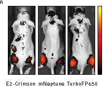
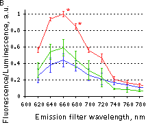
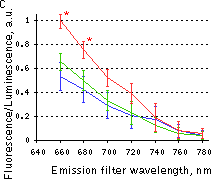
Superior performance of TurboFP650 in whole-body imaging was demonstrated using mouse xenograft model. HEK 293T cells transiently transfected with a plasmids encoding either TurboFP635, TurboFP650, NirFP, mNeptune or E2-Crimson were injected intramuscularly into the gluteal region of mice. The cells were co-transfected with firefly luciferase plasmid to normalize the transfection efficiency and total numbers of injected cells. Imaging of cell implants and quantification at various emission wavelengths showed higher fluorescence from TurboFP650 at two excitation wavelengths.
Despite its dimeric structure, TurboFP650 can be used in some fusions. However, for protein labeling applications we recommend using specially optimized monomeric TagFPs.
TurboFP650 can be used in multicolor labeling applications with blue, cyan, green, yellow, and red (orange) fluorescent dyes.
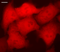 | HeLa cells transiently transfected with pTurboFP650-N vector.Widefield Leica AFLX 6000 microscope, 63x objective, after 3 days of incubation. Scale bar, 10 μm. Image from Shcherbo et al., 2010. |
|---|
 |  |  | |
|---|---|---|---|
Whole-mouse imaging with IVIS Spectrum system (Caliper).(A) Representative fluorescence reflectance images (excitation filter, 605/30 nm and emission filter, 660/20 nm) of mice injected intramuscularly with HEK 293T cells expressing E2-Crimson, mNeptune or TurboFP650. Green asterisk denotes background fluorescence in mice injected with E2-Crimson cells. The color bar indicates radiant efficiency ×10-6; minimum is 0.001, and maximum is 0.006. (B,C) Fluorescence efficiency from cell implants imaged with 570/30 nm (B) or 605/30 nm (C) excitation filters and various emission filters, normalized to photons from firefly luciferase to control for transfection efficiency and numbers of implanted cells. Means ± s.e.m. are shown (n = 6-10 per point). *P < 0.05 (Student's t-test) for TurboFP650 relative to other proteins. Red line – TurboFP650, green line – mNeptune, blue line – E2-Crimson. Images and data from Shcherbo et al., 2010. | |||
| Variant | Description | Related vector | Cat.# | |
|---|---|---|---|---|
| ||||
| Humanized TurboFP650 | TurboFP650 codon usage is optimized for high expression in mammalian cells [Haas et al., 1996], but it can be successfully expressed in many other heterological systems. Evrogen mammalian expression vectors comprising multiple cloning sites at the 5- or 3-end of TurboFP650 coding sequence allow easy generation of fusions of interest. |
|
FP731 | |
|
|
FP732 | |||
References:
- Haas J, Park EC, Seed B. Codon usage limitation in the expression of HIV-1 envelope glycoprotein. Curr Biol. 1996; 6 (3):315-24. / pmid: 8805248
- Lin MZ, McKeown MR, Ng HL, Aguilera TA, Shaner NC, Campbell RE, Adams SR, Gross LA, Ma W, Alber T, Tsien RY. Autofluorescent proteins with excitation in the optical window for intravital imaging in mammals. Chem Biol. 2009; 16 (11):1169-79. doi: 10.1016/j.chembiol.2009.10.009 / pmid: 19942140
- Shcherbo D, Shemiakina II, Ryabova AV, Luker KE, Schmidt BT, Souslova EA, Gorodnicheva TV, Strukova L, Shidlovskiy KM, Britanova OV, Zaraisky AG, Lukyanov KA, Loschenov VB, Luker GD, Chudakov DM. Near-infrared fluorescent proteins. Nat Methods. 2010; 7 (10):827-9. doi: 10.1038/nmeth.1501 / pmid: 20818379
- Strack RL, Hein B, Bhattacharyya D, Hell SW, Keenan RJ, Glick BS. A rapidly maturing far-red derivative of DsRed-Express2 for whole-cell labeling. Biochemistry. 2009; 48 (35):8279-81. doi: 10.1021/bi900870u / pmid: 19658435
|
Copyright 2002-2023 Evrogen. All rights reserved. Evrogen JSC, 16/10 Miklukho-Maklaya str., Moscow, Russia, Tel +7(495)988-4084, Fax +7(495)988-4085, e-mail:evrogen@evrogen.com |