
|
||||||||||
|
||||||||||


PhiYFP
SUPPORTRESOURCES |
|

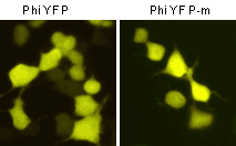 | Fluorescent microscopy of transiently transfected mammalian cells expressing Phi-Yellow proteins.
|
Suitability of Phi-Yellow proteins to generate stably transfected cells has been proven by Marinpharm company.
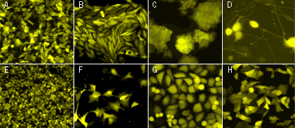 | Fluorescent microscopy of stably transfected mammalian cells expressing PhiYFP in cytosol.(A) M3 mouse melanoma; (B) T-406 human glioma; (C) PC-12 rat phaeochromocytoma cells; (D) PC-12 cells after the addition of nerve growth factor; (E) Walker 256 rat tumour cells; (F) BC3H1 cells; (G) T24 human bladder carcinoma cells; (H) T24 cells expressing destabilized variant PhiYFP-m-dest1. Images were kindly provided by Dr. Christian Petzelt (Marinpharm). |
|---|
Despite dimerization capacity, Phi-Yellow proteins demonstrate successful performance in fusions with subcellular localization signals and many cellular proteins. However, we recommend that you use TagFPs for protein labeling applications.
Important note: PhiYFP allows generation of fusions to its N-terminus, whereas PhiYFP-m is optimized to generate fusions to its C-terminus. PhiYFP can not be used to generate C-terminal fusions.
Phi-Yellow proteins can be used in multicolor labeling applications with blue, cyan, green, red, and far-red fluorescent dyes.
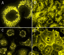 | Fluorescent microscopy of stably transfected mammalian cells expressing Phi-Yellow-tagged fusions.(A-B) Mitochondria-targeted PhiYFP in (A) 3T3 mouse fibroblasts; (B) PtK rat kangaroo cells; (C) T24 human bladder carcinoma cells expressing peroxisome-targeted PhiYFP-m; (D) PhiYFP-m fusion with β-actin in PtK rat kangaroo cells. Images were kindly provided by Dr. Christian Petzelt (Marinpharm). |
|---|
|
Copyright 2002-2023 Evrogen. All rights reserved. Evrogen JSC, 16/10 Miklukho-Maklaya str., Moscow, Russia, Tel +7(495)988-4084, Fax +7(495)988-4085, e-mail:evrogen@evrogen.com |