TagFP635


Far-red fluorescent protein TagFP635

- Bright far-red fluorescence
- Monomer with successful performance in fusions
- High photostability
- Fluorescent signal is easily distinguished from background fluorescence
- Recommended for protein labeling

TagFP635 (scientific name mKate) is a monomeric far-red fluorescent protein generated from the wild-type RFP from sea anemone Entacmaea quadricolor [Shcherbo et al., 2007]. It possesses bright fluorescence with excitation/emission maxima at 588 and 635 nm, respectively.
TagFP635 is 6.4-fold brighter than mPlum, 1.25-fold brighter and much more photostable than mRaspberry [Shcherbo et al., 2007; Shaner et al., 2004]. This makes it the best monomeric far-red fluorescent protein available so far.
TagFP635 is mainly intended for protein labeling. It fluorescence allows easy and reliable separation from standard green fluorescent labels in dual-color high-throughput assays. TagFP635 can also be used for cell and organelle labeling and for tracking the promoter activity; however, TurboFP635 is preferable for these applications as it is brighter and more pH-stable than TagFP635.

Main properties

TagFP635 spectra

TagFP635 normalized excitation (thin line) and emission (thick line) spectra.

Download TagFP635 spectra (xls)

CHARACTERISTIC
*Brightness is a product of extinction coefficient and quantum yield, divided by 1000.
Molecular weight, kDa27
Polypeptide length, aa237
Fluorescence colorfar-red
Excitation maximum, nm635
Emission maximum, nm588
Quantum yield0.33
Extinction coefficient, M-1cm-145000
Brightness*14.9
Brightness, % of EGFP45
pKa6.0
Structuremonomer
Aggregationno
Maturation rate at 37°Cfast
Photostabilityhigh
Main advantagesmonomeric far-red fluorescent protein, high photostability

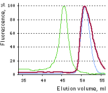
Gel-filtration of TurboGFP (dimer, green line), EGFP (monomer, blue line), and TagFP635 (monomer, red line).

Image from Shcherbo et al., 2007.

Recommended filter sets and antibodies

TagFP635 can be recognized using Anti-tRFP antibody (Cat.# AB231-AB232) available from Evrogen.

Recommended Omega Optical filter sets are QMAX-Red and XF102-2. TagFP635 can also be detected using Texas Red filter sets or similar.

Performance and use

TagFP635 can be easily expressed and detected in a wide range of organisms. It becomes clearly detectable in mammalian cells as early as within 12-14 hrs after transfection. No cell toxic effects and visible protein aggregation are observed.
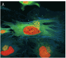
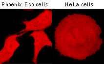
TagFP use for cell labeling.

Transiently transfected mamalian cells expressing TagFP635.
Image of Phoenix Eco cells is from Shcherbo et al., 2007; image of a HeLa cell was kindly provided by Michael W. Davidson (Florida State University).

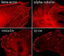
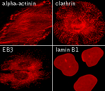
TagFP635 performance in fusions has been demonstrated in α-actinin, vinculin, zyxin, β-actin, α-tubulin, and other models.

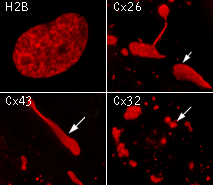
TagFP635 use for protein labeling in mammalian cells.

Microscopic images of HeLa cells transiently transfected with TagFP635-tagged fusions. Arrows indicate gap junction.
Images were kindly provided by Michael W. Davidson (Florida State University).

TagFP635 can be used in multicolor labeling applications with blue, cyan, green, yellow, and red (orange) fluorescent proteins.

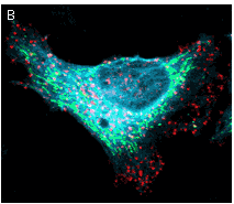
TagFP635 use in multicolor labeling of mammalian cells.

(A) TagCFP-tagged β-actin (cyan), TagYFP-tagged α-tubulin (green), TagFP635-H2B fusion (red), and Golgi-targeted TagRFP (yellow); (B) TagCFP-tagged α-tubulin (cyan), TagFP635-clathrin fusion (red), mitochondria-targeted TagYFP (green). Images were kindly provided by Michael W. Davidson (Florida State University).

Available variants and fusions

TagFP635 codon usage is optimized for high expression in mammalian cells [Haas et al., 1996], but it can be successfully expressed in many other heterological systems. Evrogen mammalian expression vectors comprising multiple cloning sites at the 5'- or 3'-end of TagFP635 coding sequence allow easy generation of fusions of interest.

TagFP635-actinin fusion: Human α-actinin is fused to the TagFP635 N-terminus. When expressed in mammalian cells, this fusion provides red fluorescent labeling of α-actinin in living cells.

TagFP635-tubulin fusion: Human α-tubulin is fused to the TagFP635 C-terminus. When expressed in mammalian cells, this fusion provides red fluorescent labeling of α-tubulin in living cells.

TagFP635-clathrin fusion: Human clathrin light chain LCB is fused to the TagFP635 C-terminus. When expressed in mammalian cells, this fusion provides red fluorescent labeling of clathrin light chain LCB in living cells.

TagFP635-actin fusion: Human β-actin is linked to the TagFP635 C-terminus. When expressed in mammalian cells, this fusion provides red fluorescent labeling of β-actin in living cells.

TagFP635-EB3 fusion: Human microtubule-associated EB3 protein is fused to the TagFP635 N-terminus. When expressed in mammalian cells, this fusion provides red fluorescent labeling of EB3 protein in living cells.

TagFP635-vinculin fusion: Human vinculin is fused to the TagFP635 C-terminus. When expressed in mammalian cells, this fusion provides red fluorescent labeling of vinculin in living cells.

TagFP635-zyxin fusion: Human zyxin is fused to the TagFP635 N-terminus. When expressed in mammalian cells, this fusion provides red fluorescent labeling of zyxin in living cells.

TagFP635-Cx26 fusion: Rat connexin 26 is fused to the TagFP635 N-terminus. When expressed in mammalian cells, this fusion provides red fluorescent labeling of connexin 26 in living cells.

TagFP635-Cx32 fusion: Human connexin 32 is fused to the TagFP635 N-terminus. When expressed in mammalian cells, this fusion provides red fluorescent labeling of connexin 32 in living cells.

TagFP635-Cx43 fusion: Rat connexin 43 is fused to the TagFP635 N-terminus. When expressed in mammalian cells, this fusion provides red fluorescent labeling of connexin 43 in living cells.

TagFP635-H2B fusion: Human histone H2B is fused to the TagFP635 N-terminus. When expressed in mammalian cells, this fusion provides red fluorescent labeling of histone H2B in living cells.

TagFP635-laminB1 fusion: Human lamin B1 is fused to the TagFP635 C-terminus. When expressed in mammalian cells, this fusion provides red fluorescent labeling of lamin B1 in living cells.

References:

  • Haas J, Park EC, Seed B. Codon usage limitation in the expression of HIV-1 envelope glycoprotein. Curr Biol. 1996; 6 (3):315-24. / pmid: 8805248
  • Shaner NC, Campbell RE, Steinbach PA, Giepmans BN, Palmer AE, Tsien RY. Improved monomeric red, orange and yellow fluorescent proteins derived from Discosoma sp. red fluorescent protein. Nat Biotechnol. 2004; 22 (12):1567-72. / pmid: 15558047
  • Shcherbo D, Merzlyak EM, Chepurnykh TV, Fradkov AF, Ermakova GV, Solovieva EA, Lukyanov KA, Bogdanova EA, Zaraisky AG, Lukyanov S, Chudakov DM. Bright far-red fluorescent protein for whole-body imaging. Nat Methods. 2007; 4 (9):741-6. / pmid: 17721542
Copyright 2002-2008 Evrogen. All rights reserved.
Evrogen JSC, 16/10 Miklukho-Maklaya str., Moscow, Russia, +7(495)4298020, e-mail:evrogen@evrogen.com