|
Red (orange) fluorescent protein TurboRFP
- Super bright red (orange) fluorescence
- Fast maturation, high pH-stability
- Recommended for gene expression analysis and cell and organelle labeling
 |
TurboRFP is a red (orange) fluorescent protein (excitation/emission maxima are 553 and 574 nm, respectively) derived from sea anemone Entacmaea quadricolor [Merzlyak et al., 2007]. Possessing high photostability and pH stability, TurboRFP is more than twice brighter than DsRed2. Fast TurboRFP maturation makes it clearly detectable in mammalian cells as early as within 8-10 hrs after transfection.
TurboRFP is mainly intended for applications where fast appearance of bright fluorescence is crucial. It is specially recommended for cell and organelle labeling and tracking the promoter activity.
|
Main properties
TurboRFP normalized excitation (thin line) and emission (thick line) spectra.
Spectra viewer tool
Download TurboRFP spectra (xls)
| | CHARACTERISTIC | |
|---|
* Brightness is a product of extinction coefficient and quantum yield, divided by 1000.
| | Molecular weight, kDa | 26.1 | | Polypeptide length, aa | 231 | | Fluorescence color | red (orange) | | Excitation maximum, nm | 553 | | Emission maximum, nm | 574 | | Quantum yield | 0.67 | | Extinction coefficient, M-1cm-1 | 92 000 | | Brightness* | 61.6 | | Brightness, % of EGFP | 187 | | pKa | 4.4 | | Structure | dimer | | Aggregation | no | | Maturation rate at 37°C | super fast | | Photostability | high | | Cell toxicity | not observed | | Main advantages | superbright and fast-maturing red fluorescent protein | | Possible limitations | dimer, limited applicability for fusions generation |
|
|---|
Recommended filter sets and antibodies
TurboRFP can be recognized using Anti-tRFP antibody (Cat.# AB233) available from Evrogen.
Recommended Omega Optical filter sets are QMAX-Yellow, XF108-2, XF101-2, and XF111-2. TurboRFP can also be detected using TRITC filter set or similar.
Performance and use
TurboRFP can be expressed and detected in a wide range of organisms. Mammalian cells transiently transfected with TurboRFP expression vectors produce bright fluorescence in 8-10 hrs after transfection. No cytotoxic effects or visible protein aggregation are observed.
Despite its dimeric structure, TurboRFP performs well in some fusions. However, for protein labeling applications we recommend using specially optimized monomeric TagFPs.
TurboRFP can be used in multicolor labeling applications with blue, cyan, green, yellow, and far-red fluorescent dyes.
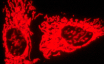
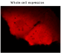 | 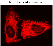 | TurboRFP use for cell and organelle labeling.
Fluorescent microscopy of mammalian cells expressing cytoplasmic TurboRFP and TurboRFP fusion with mitochondrial targeting signal. Images made from HeLa cells 24 hrs after transfection.
|
|---|
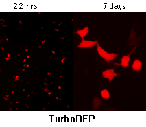
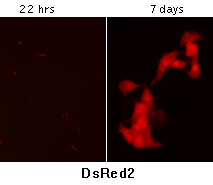
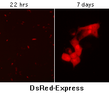
TurboRFP maturation kinetics: TurboRFP matures noticeably faster than other red fluorescent proteins. To compare maturation of TurboRFP and DsRed-related proteins, HeLa cells were transiently transfected with mammalian expression vectors comprising TurboRFP, DsRed2, or DsRed-Express fluorescent proteins under the control of CMV promoter. The DNA concentrations were equalized before transfection. Cells were photographed using fluorescent microscope after different periods of cultivation. Faster appearance of bright fluorescence was detected in the case of TurboRFP. In addition, unlike DsRed-related proteins, no abnormal Golgi-like localization of TurboRFP was observed within 7 days after transfection.
| Characteristic | TurboRFP | DsRed2** | DsRed-Express** |
|---|
* Brightness is a product of extinction coefficient and quantum yield, divided by 1000.
** DsRed2 and DsRed-Express characteristics available from literature sources are shown. Literature information regarding DsRed2 extinction coefficient and brightness differs from our data shown in parentheses. | | Fluorescence color | red (orange) | red (orange) | red (orange) | | Excitation max (nm) | 553 | 563 | 557 | | Emission max (nm) | 574 | 582 | 579 | | Quantum yield | 0.67 | 0.55 | 0.4 | | Extinction coefficient (M-1cm-1) | 92 000 | 43 800 (65 000) | 30 100 | | Brightness* | 61.6 | 24.1 (35.8) | 12.6 | | pKa | 4.4 | 4.5 | no data | | Molecular weight | 26.1 kDa | 25.8 kDa | 25.7 kDa | | Structure | dimer | tetramer | tetramer | Detection in mammalian cells
(hrs after transfection) | 8-12 | 36-48 | 8-12 |
Available variants and fusions
| Variant | Description | Related vector | Cat.# | Click for image |
|---|
|
|
Humanized TurboRFP
|
TurboRFP codon usage is optimized for high expression in mammalian cells [Haas et al., 1996], but it can be successfully expressed in many other heterological systems.
|
pTurboRFP-C
|
FP231
|
|
pTurboRFP-N
|
FP232
|
|
pTurboRFP-B
|
FP233
|
|
pTurboRFP-PRL
|
FP235
|
|
TurboRFP-mito fusion
|
A mitochondrial targeting sequence (MTS) is fused to the TurboRFP N-terminus. MTS was derived from the subunit VIII of human cytochrome C oxidase [Rizzuto et al., 1989; Rizzuto et al., 1995]. When expressed in mammalian cells, this variant provides red (orange) fluorescent labeling of mitochondria.
|
pTurboRFP-mito
|
FP237
|
|
References:
-
Haas J, Park EC, Seed B.
Codon usage limitation in the expression of HIV-1 envelope glycoprotein.
Curr Biol. 1996; 6 (3):315-24. / pmid: 8805248
-
Merzlyak EM, Goedhart J, Shcherbo D, Bulina ME, Shcheglov AS, Fradkov AF, Gaintzeva A, Lukyanov KA, Lukyanov S, Gadella TW, Chudakov DM.
Bright monomeric red fluorescent protein with an extended fluorescence lifetime.
Nat Methods. 2007; 4 (7):555-7. / pmid: 17572680
-
Rizzuto R, Brini M, Pizzo P, Murgia M, Pozzan T.
Chimeric green fluorescent protein as a tool for visualizing subcellular organelles in living cells.
Curr Biol. 1995; 5 (6):635-42. / pmid: 7552174
-
Rizzuto R, Nakase H, Darras B, Francke U, Fabrizi GM, Mengel T, Walsh F, Kadenbach B, DiMauro S, Schon EA.
A gene specifying subunit VIII of human cytochrome c oxidase is localized to chromosome 11 and is expressed in both muscle and non-muscle tissues.
J Biol Chem. 1989; 264 (18):10595-600. / pmid: 2543673
|